Why Accurate Radiographic Scaling Is Essential for Orthopedic Templating—and How Akucal Outperforms the Competition

Why Accurate Radiographic Scaling Is Essential for Orthopedic Templating—and How Akucal Outperforms the Competition

When it comes to orthopedic templating, accuracy is paramount. A tiny miscalculation in implant sizing can lead to complications, revision surgeries, and skyrocketing costs. In fact, radiographic scaling is arguably the most important step of the templating process—if you’re not working from a precisely scaled image, every other aspect of surgical planning is built on uncertain ground. Below, we’ll explore why accurate scaling is so critical, how it saves both time and money, and why the Akucal stands out as the best investment an orthopedic practice can make.

1. The Critical Importance of Accurate Scaling

Precision Impacts Outcomes

  • Choosing the Correct Implant
    Implant mismatches don’t just risk patient outcomes—they can cost a fortune in revised procedures and extended recovery times. Accurate scaling is your insurance policy against these avoidable pitfalls.

  • Reducing Revisions & Complications
    A revision surgery can cost tens of thousands of dollars, not to mention the extra strain on patients. By ensuring you begin with precisely scaled images, you drastically lower the risk of post-op surprises and the need for corrective procedures.

Scaling as the Foundation of Templating

Whether you’re planning a total hip replacement or fixing a complex fracture, the success of the surgery depends on having the right measurements. Many facilities invest millions in advanced surgical technology but overlook the simple step that could make or break their outcomes: obtaining an accurately scaled radiograph.

2. Akucal: The Pinnacle of Radiographic Scaling

While the importance of scaling may be clear, not all markers are created equal. The Akucal was custom-engineered to meet the high stakes of orthopedic templating, featuring:

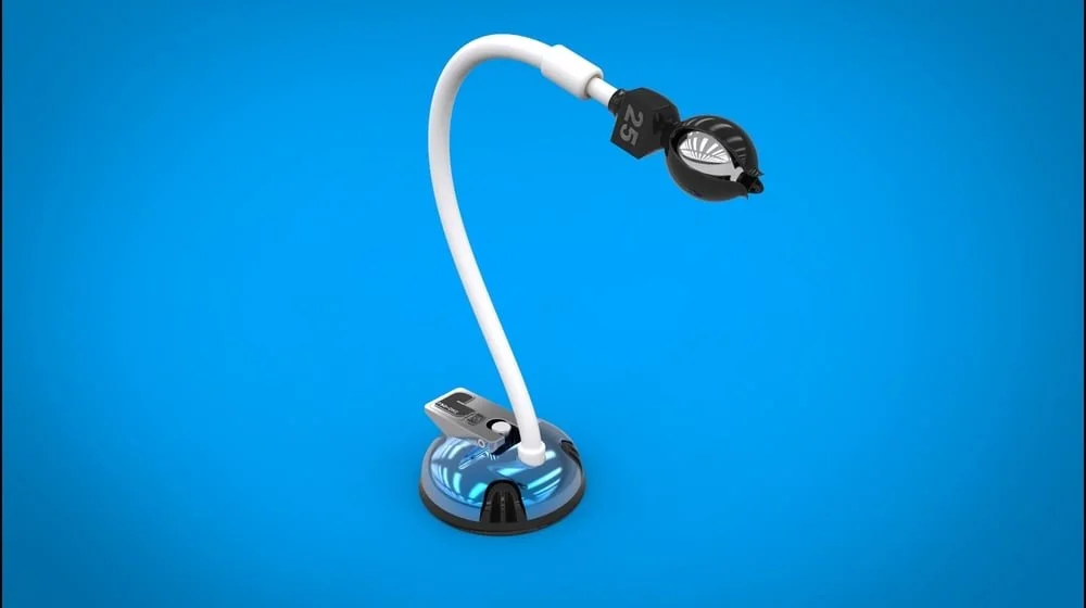
CNC’d Aluminum Suction Base

  • Unrivaled Strength
    Each Akucal suction base is CNC-machined from a solid block of aluminum, taking hours to craft. The result? A robust, secure seal that won’t slip—even under the most demanding conditions.

  • No More Shifting Mid-Procedure
    Say goodbye to wobbly, plastic bases that compromise your accuracy at the worst possible moment.

Fully Covered, Flexible Gooseneck Arm

  • Easy to Clean
    With no exposed crevices, debris can’t hide, allowing for fast and thorough sanitization.

  • Smooth & Quiet Adjustment
    Unlike squeaky coolant hoses used by many competitors, the Akucal’s covered gooseneck is designed specifically for clinical use.

Integrated Marker Size Indicators

  • Mandatory for True Accuracy
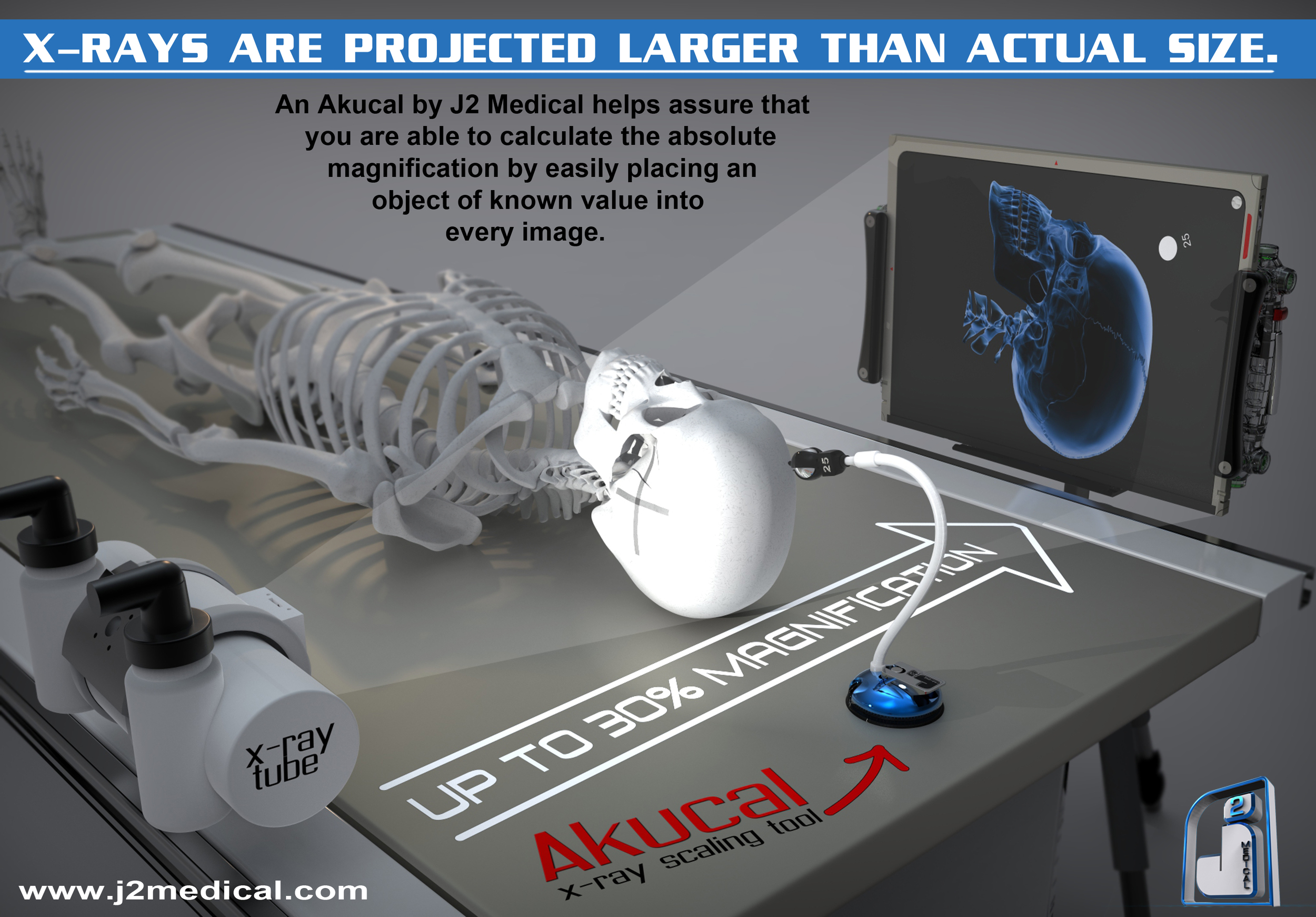
    Built-in lead markings ensure the marker’s size is visible on every radiograph, making it easy to confirm calibration. This single feature is a game-changer for orthopedic software providers, who rely on consistent, accurate reference points for digital templating.

Modular “Ballclaw” Tip

  • Quick Removal, No Tools Needed
    The entire tip detaches in seconds, simplifying cleaning or replacement.

  • Streamlined Maintenance
    Easily swap the tip if it becomes damaged or needs sanitizing, without halting your workflow.

Versatile Accessories

  • Extra Markers & Ballclaws – Swap them out on the fly.

  • “Undercover” Shield – Absorbs radiation in bariatric cases to prevent marker burnout.

  • Rigid Base – Suction your Akucal onto padded or textured surfaces with ease.

3. How Competitors Fall Short

While the Akucal is thoughtfully engineered from the ground up, many competitors rely on OEM parts pieced together—leading to inevitable issues:

  1. Cheap Plastic Bases
    Prone to cracking or losing suction at critical moments, compromising your carefully planned scaling.

  2. Squeaky Industrial Coolant Hose
    Hard-to-clean segmented hoses gather debris, creating infection control concerns and diminishing the professional feel of your radiography setup.

  3. Hollow Markers, No Size Indicators
    Hollow markers risk burning out under higher beam settings, especially for bariatric patients. Worse yet, the lack of size indicators means there’s no reliable reference in your images, negating any confidence in digital templates.

4. The Financial and Clinical ROI

Saves Time and Money—Instantly

  • One Surgery = Instant Payback
    If a single procedure is optimized by accurate scaling—leading to the correct implant size, less OR time, and fewer complications—the Akucal essentially pays for itself.

  • Fewer Revisions, Less OR Disruption
    Revision surgeries are expensive and time-consuming. By reducing the chances of implant miscalculation, you protect both patient well-being and your bottom line.

Boosts Efficiency in Workflow

  • Faster, More Confident Templating
    Consistent scaling markers empower surgeons and radiologists to trust their measurements from day one, speeding up planning and reducing second-guessing.

  • Optimized for Orthopedic Software
    Clear lead markings enhance digital templating, making it straightforward for orthopedic software providers to integrate scaling data seamlessly.

5. Conclusion: The Smartest Investment in Orthopedic Imaging

From selecting implants to preventing costly revisions, radiographic scaling is the bedrock of successful orthopedic surgery. The Akucal has taken a seemingly simple process and elevated it into a high-precision science—one that helps thousands of surgeons worldwide achieve better outcomes, save time, and save money. With its CNC’d aluminum suction base, fully covered gooseneck arm, built-in lead size indicators, and modular Ballclaw tip, the Akucal outshines its competitors in every critical dimension.

When accurate scaling is paramount—and when even a single error can lead to significant financial and clinical repercussions—why settle for less than the best? By investing in the Akucal, you’re not just buying a marker. You’re investing in a purpose-built solution that ensures each procedure is set up for success. After all, it only takes one properly scaled surgery to realize the true value—and watch the Akucal pay for itself the very first time you use it.

Ortho Surgeons: Would You Invest $400 to Save $2.5 Million

Ortho Surgeons: Would You Invest $400 to Save $2.5 Million

In the world of orthopedic surgery, precision, efficiency, and cost-effectiveness are paramount. Surgeons constantly seek innovative solutions that enhance patient outcomes while optimizing their practice's financial health. Today, we pose a question that could reshape the way ortho surgeons view their investments. Would you spend $400 to save a staggering $2.5 million? Let's delve into this intriguing proposition.

**The Power of $400: Introducing the Akucal**

First, let's meet the star of the show – the Akucal Image Scaling Tool. Priced at a mere $400 per unit, this revolutionary tool is designed to eliminate guesswork in image scaling. By precisely determining magnification factors, it ensures that ortho surgeons choose the right implant sizes with confidence. This translates to fewer adjustments during surgeries and a significant reduction in hardware wastage.

**The Cost Savings Potential: A Closer Look**

Now, let's crunch the numbers. Imagine a hospital or orthopedic practice performing 20 surgeries a day. Over the course of a year, that's 5,000 surgeries. Without the Akucal, the average hardware wastage cost per surgery is $1,000. This would result in an astonishing $5,000,000 in hardware wastage costs.

However, with the Akucal in place, let's assume a conservative 50% reduction in hardware wastage costs. This means saving $500 per surgery, resulting in a total savings of $2,500,000 over the year. And remember, the Akucal's initial investment cost is just $400 per unit.

**The Question of Investment: $400 for $2.5 Million in Savings?**

Here's the heart of the matter: Would you be willing to invest $400 in an Akucal unit to save $2.5 million in hardware wastage costs over the course of a year? The answer seems almost self-evident.

By adopting the Akucal, ortho surgeons not only safeguard their practice's financial health but also enhance precision, efficiency, and patient outcomes. It's not just an investment; it's a financial transformation that empowers surgeons to provide the highest level of care while optimizing costs.

**Conclusion: The Akucal - A Wise Investment**

In an era where every dollar counts, the Akucal proves to be a brilliant investment for ortho surgeons and healthcare facilities. It's a tool that doesn't just save money; it unlocks the potential for unparalleled financial efficiency without compromising patient care.

So, ortho surgeons, the question remains: Would you spend $400 to save $2.5 million? The choice seems clear, and the Akucal is here to help you make it.

If you're ready to explore the transformative potential of the Akucal in your practice, don't hesitate to reach out. The future of orthopedic surgery is precision, efficiency, and cost-effectiveness, and the Akucal can help you achieve just that.

Unlocking Efficiency and Precision in Ortho: The Vital Role of Akucals in Modern Workflows

Unlocking Efficiency and Precision in Ortho: The Vital Role of Akucals in Modern Workflows

Scaling an x-ray with an Akucal (also known as a calibration marker) is a crucial step in joint replacement surgery because it allows the surgeon to accurately determine the size of the implant needed for the patient.

During joint replacement surgery, the surgeon removes the damaged joint and replaces it with an artificial implant. The implant must be the right size and shape to fit the patient's anatomy and provide proper function.

To determine the appropriate size of the implant, the surgeon uses pre-operative imaging such as X-rays. However, these images can be distorted due to differences in magnification and positioning, which can lead to inaccurate measurements.

Using an Akucal allows the surgeon to account for any distortion in the imaging and accurately scale the image. The Akucal is a small metal marker placed in the same plane as the joint being replaced, and it has known dimensions that can be used to accurately measure the size of the implant needed.

Without scaling the X-ray with an Akucal, there is a risk of using an implant that is too large or too small for the patient, which can lead to improper function, instability, and premature wear and failure of the implant.

Therefore, scaling an x-ray with an Akucal is an essential step in joint replacement surgery to ensure proper sizing and placement of the implant, and ultimately a successful outcome for the patient.


Elevating Hip Replacement Surgeries: The Akucal's Cutting-Edge Advantage

Elevating Hip Replacement Surgeries: The Akucal's Cutting-Edge Advantage

Introduction:

In the dynamic world of medical advancements, innovation continues to shape the way surgeries are performed. Among these advancements, the Akucal emerges as a trailblazing tool that's set to redefine hip replacement procedures. Let's take a quick dive into the transformative power of the Akucal and its potential to revolutionize the landscape of surgical precision.

Precision Perfected: The Akucal's Game-Changing Technology

Imagine a scenario where every hip replacement surgery is carried out with absolute precision. The Akucal makes this a reality. At the heart of its design lies the ability to calculate magnification factors with unparalleled accuracy. Gone are the days of estimation and guesswork. With the Akucal, surgeons gain real-time measurements that ensure a perfect fit every time, reducing the need for adjustments and reworks.

Efficiency Unleashed: Streamlined Workflow and Surgeon Confidence

Surgical procedures are intricate dances that require every step to be choreographed flawlessly. The Akucal becomes the choreographer, allowing surgeons to glide through surgeries with newfound efficiency. By eliminating the need for guesswork, surgeons can focus on delivering exceptional care. The result? Surgeon confidence soars, and patients benefit from streamlined, high-quality procedures.

Cost Savings in Focus: A Financial Transformation

It's no secret that surgical procedures come with costs, both in terms of resources and time. The Akucal addresses a significant cost factor – hardware wastage. With precise measurements, wastage due to inaccurate fittings becomes a thing of the past. This not only translates to cost savings but also to the ability to allocate resources where they matter most – patient care and further advancements.

Patient-Centric Approach: Improved Outcomes and Satisfaction

At the heart of every medical advancement lies the goal of improving patient outcomes. The Akucal delivers on this promise by ensuring implants fit perfectly. This means improved functionality, quicker recovery times, and ultimately, happier patients. With the Akucal, patient satisfaction becomes an integral part of the surgical equation.

Embrace the Future with the Akucal

As the medical landscape evolves, embracing innovative technologies becomes paramount. The Akucal isn't just a tool; it's a beacon of progress. Surgeons equipped with the Akucal are at the forefront of change, ready to transform how hip replacement surgeries are performed. Step into the future of surgical excellence – the Akucal way.

Conclusion:

The Akucal isn't just an advancement; it's a revolution. In a world where precision, efficiency, and cost-effectiveness are paramount, the Akucal stands tall. It redefines surgical procedures, ensuring every patient receives the utmost care with minimal wastage. As we move forward in the realm of medical innovation, the Akucal becomes a shining example of how technology and healthcare can intertwine to create a brighter future for all.

Simple Calculator For X-ray Magnification after using an Akucal

Simple Calculator For X-ray Magnification after using an Akucal

X-ray Magnification Calculator

X-ray Magnification Calculator


To use the calculator:

  1. Input the actual marker size (for instance, 25mm).

  2. Input the measured marker size from the x-ray image.

  3. Click on "Calculate Magnification" to get the magnification factor and percentage.

Scaling Calculator

Scaling Calculator


In this calculator:

  1. Users will input their original measurement.

  2. Users will also input the magnification percentage (e.g., 10 for 10% magnification).

  3. Clicking the "Calculate Scaled Measurement" button will give them the scaled-up measurement based on the entered magnification percentage.

A Statement to Orthopedic Templating Software Companies

A Statement to Orthopedic Templating Software Companies

In the dynamic realm of orthopedic surgery and diagnostics, the importance of precision cannot be understated. Over the past 17 years, J2 Medical's Akucal has set a benchmark in the industry, scaling millions of X-rays for tens of thousands of esteemed radiologists and surgeons across the globe. Our unwavering commitment to ensuring every X-ray possesses an accurate scale factor embodies the future we envision for the orthopedic community.

To our peers and innovators in the orthopedic templating software landscape:

We recognize the transformative work you're doing, and we are inspired by the technological advancements we've witnessed in recent years. As we continue to champion the cause of standardized scaling through Akucal, we believe in the immense potential of collaboration and mutual growth.

In an era where technology is revolutionizing medical care, it's our collective responsibility to ensure that tools are not just advanced but also accurate, reliable, and user-friendly. By aligning our endeavors, we can elevate the quality of care, contribute to better patient outcomes, and shape the future of orthopedic imaging and templating.

We are open to dialogue, partnerships, and collaborative ventures that can further the shared goal of excellence in orthopedic care. The future is bright, and together, we can make it even brighter.

Here's to innovation, collaboration, and a future where every X-ray is scaled to perfection.

J2 Medical, LP

www.j2medical.com

From Akucal to Scale: Determine X-ray Magnification without using software

From Akucal to Scale: Determine X-ray Magnification without using software

X-ray imaging has been pivotal in advancing medical diagnostics. But with its widespread use, there's an element often overlooked: magnification. In this post, we'll demystify the concept of X-ray magnification using a standard Akucal 25mm sphere marker.

---

Understanding the Basics

Magnification in X-rays arises from the unique geometry between the X-ray source, the object under examination, and the image receptor (like a digital detector or film). This magnification can be influenced by multiple factors, including the distance between the X-ray source and the patient and between the patient and the detector.

---

Why Use a 25mm Sphere Marker?

A radiopaque marker provides a known reference within the X-ray image, allowing clinicians and technicians to accurately calculate and understand the degree of magnification present. The 25mm sphere marker is commonly used because of its standardized size, which makes calculations straightforward.

---

Calculating Magnification: A Simple Formula

To determine the magnification using the 25mm sphere, use the following formula:

Magnification Factor (MF) = Size of the sphere on the image / 25 mm (actual size)

To deduce the magnification percentage:

Magnification Percentage = (MF - 1) x 100%

---

Illustrative Examples

- Example 1: For a 25mm sphere appearing as 27.5mm on the X-ray image, the magnification is 10%.

- Example 2: If the 25mm sphere measures 32.5mm on the X-ray image, the magnification becomes 30%.

- Example 3: A sphere that measures 40mm on the X-ray image has undergone a magnification of 60%.

Conclusion

Understanding X-ray magnification is vital, especially in orthopedic procedures where precision matters. With the 25mm sphere marker, professionals can decode this magnification, ensuring accuracy and better patient outcomes. As the realm of radiography continues to evolve, having foundational knowledge about elements like magnification remains crucial.

Why Orthopedic Templating Software is Inaccurate

Why Orthopedic Templating Software is Inaccurate

Simply put… It’s not the software! You have to properly consider the magnification factor.

Scaling an x-ray with an Akucal (also known as a calibration marker) is a crucial step in joint replacement surgery because it allows the surgeon to accurately determine the size of the implant needed for the patient.

During joint replacement surgery, the surgeon removes the damaged joint and replaces it with an artificial implant. The implant must be the right size and shape to fit the patient's anatomy and provide proper function.

To determine the appropriate size of the implant, the surgeon uses pre-operative imaging such as X-rays. However, these images can be distorted due to differences in magnification and positioning, which can lead to inaccurate measurements.

Using an Akucal allows the surgeon to account for any distortion in the imaging and accurately scale the image. The Akucal is an ingenious tool that is placed in the same plane as the joint being replaced, and it has known dimensions that can be used to accurately measure the size of the implant needed.

Without scaling the X-ray with an Akucal, there is a risk of using an implant that is too large or too small for the patient, which can lead to improper function, instability, and premature wear and failure of the implant.

Therefore, scaling an x-ray with an Akucal is an essential step in joint replacement surgery to ensure proper sizing and placement of the implant, and ultimately a successful outcome for the patient.

Scaling Marker Position Determines the Accuracy of Digital Templating for Total Hip Arthroplasty

Scaling Marker Position Determines the Accuracy of Digital Templating for Total Hip Arthroplasty

REPOST from PubMed. Full study here: 29398996

. 2018 Feb;14(1):55-59.

doi: 10.1007/s11420-017-9578-0. Epub 2017 Sep 12.

Scaling Marker Position Determines the Accuracy of Digital Templating for Total Hip Arthroplasty

Austin J Ramme 1Nina D Fisher 1Jonathan Egol 1Gregory Chang 2Jonathan M Vigdorchik 1

Affiliations expand

Free PMC article

Abstract

Background: Digital templating systems foster patient-specific measurements for preoperative planning.

Questions/purposes: We aim (1) to verify the accuracy of a templating system, (2) to describe the effects of scaling marker position on the accuracy of digital templating of the hip, and (3) to provide a practical guide for scaling marker position using patient body mass index (BMI).

Methods: A scaling sphere was placed in five positions along the anterior-posterior axis of an acetabular implant and pelvis phantom, and x-rays were obtained. Each radiograph was templated for the acetabular component and recorded. A retrospective review identified CT scans of preoperative hip arthroplasty cases. The center of the greater trochanter was calculated from these CT scans as the percent distance from the anterior thigh and recorded with the patient's BMI.

Results: By centering the scaling sphere on the acetabular component, an accurate cup size was achieved. A difference of 3.5 cm in sphere placement resulted in a full cup size magnification error. Positioning the scaling sphere at the level of the pubic symphysis resulted in a difference of four cup sizes. This patient population had an average BMI of 28.72 kg/m2 (standard deviation 6.26 kg/m2) and an average position of the center of the greater trochanter of 51% (standard deviation of 6%) from the anterior surface of thigh.

Conclusions: Digital templating relies on scaling marker position to accurately estimate implant size. Based on the findings in this study, scaling markers for hip imaging should be placed laterally, mid-thigh in the anterior-posterior direction for patients with a BMI between 25 and 40 kg/m2. If abnormal hip anatomy or extremes of BMI are discovered, then scaling sphere positioning should be optimized on a case-by-case basis. Digital templating systems for total hip arthroplasty must use precisely placed scaling markers at the level of the hip joint to allow for accurate implant size estimation.

Keywords: digital templating; radiographs; scaling; surgical planning; total hip arthroplasty.